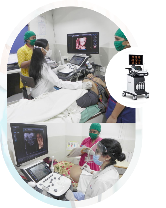
3D 4D Sonography Services
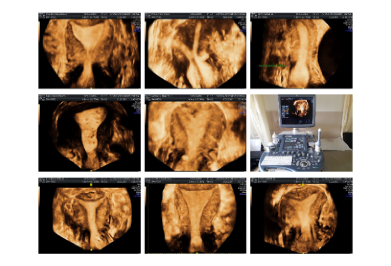
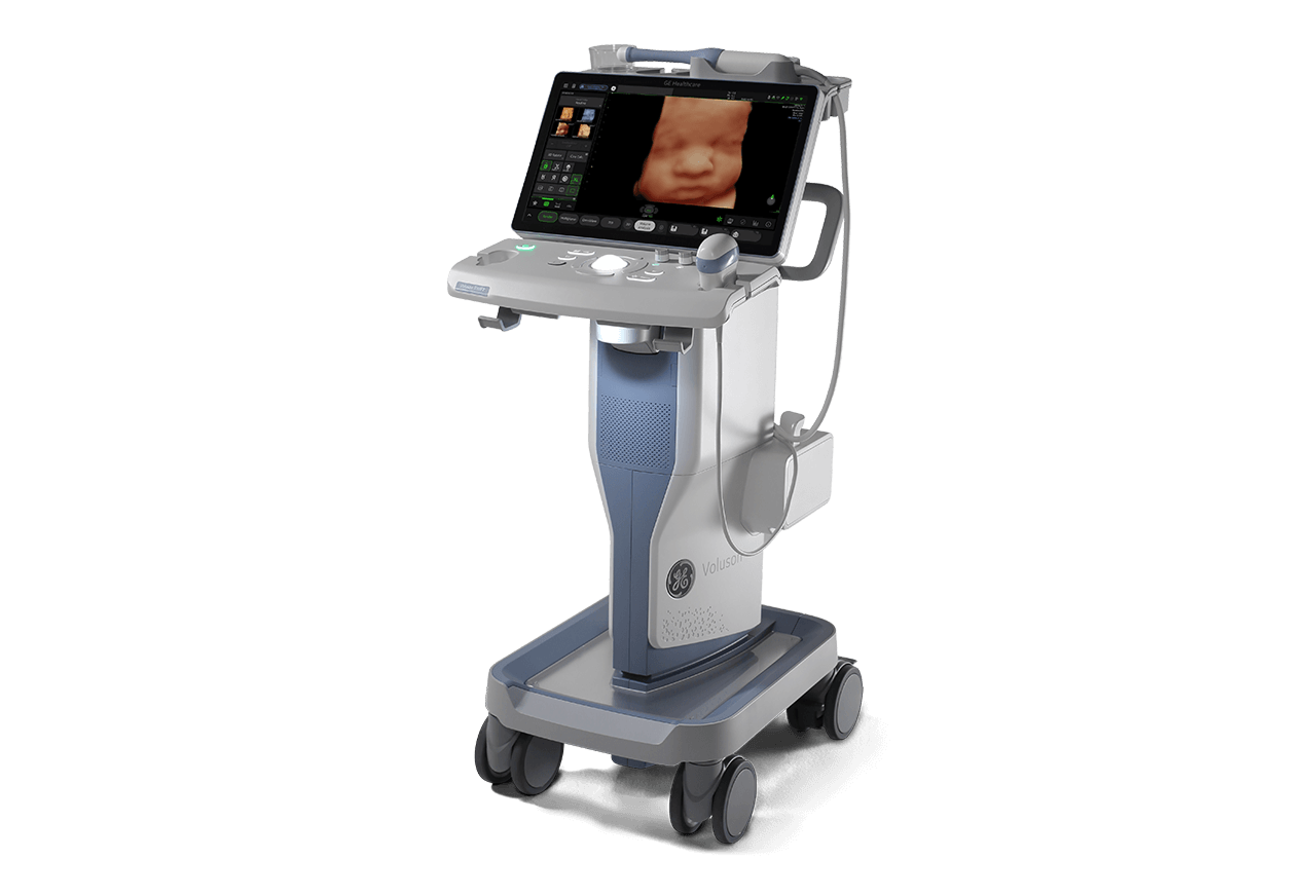
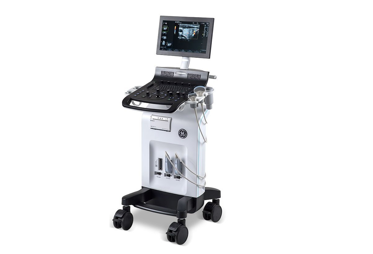
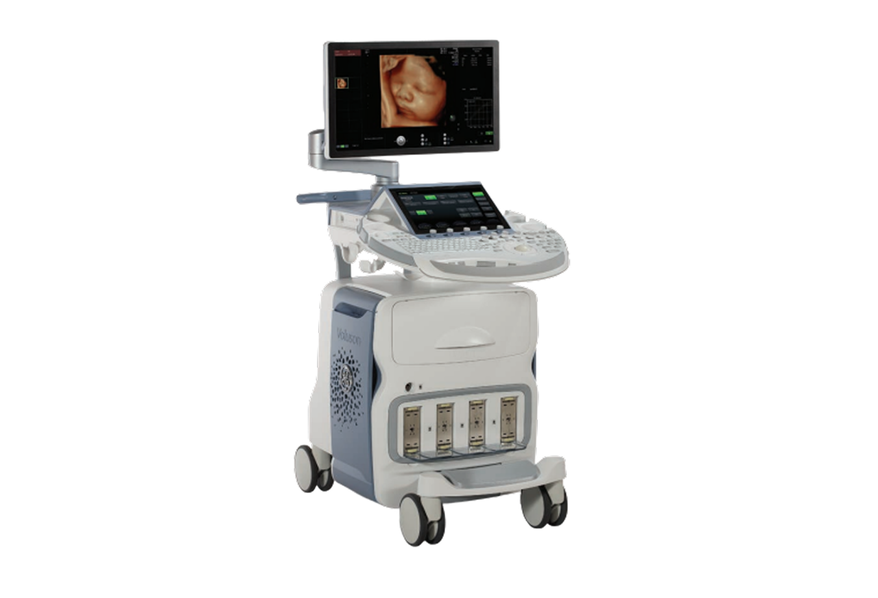
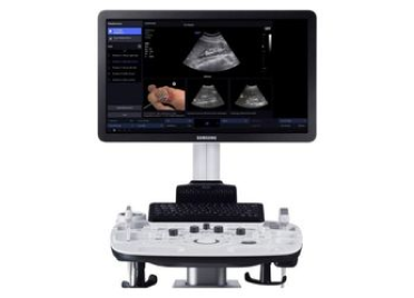
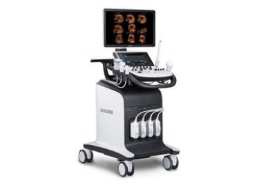
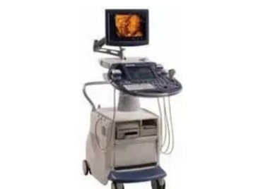
Our USG department equipped with five state of the art modern ULTRASOUND MACHINES (3D/4D/5D).
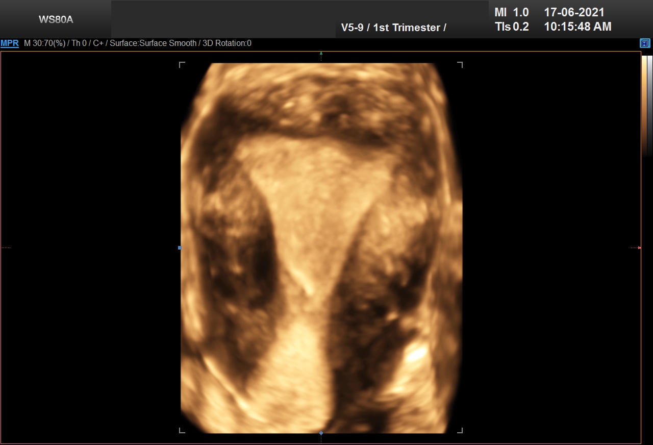
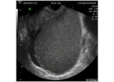
Ultrasound provides highest resolution image and helps to diagnose anatomical defects in the foetus.
- HS30 SAMSUNG
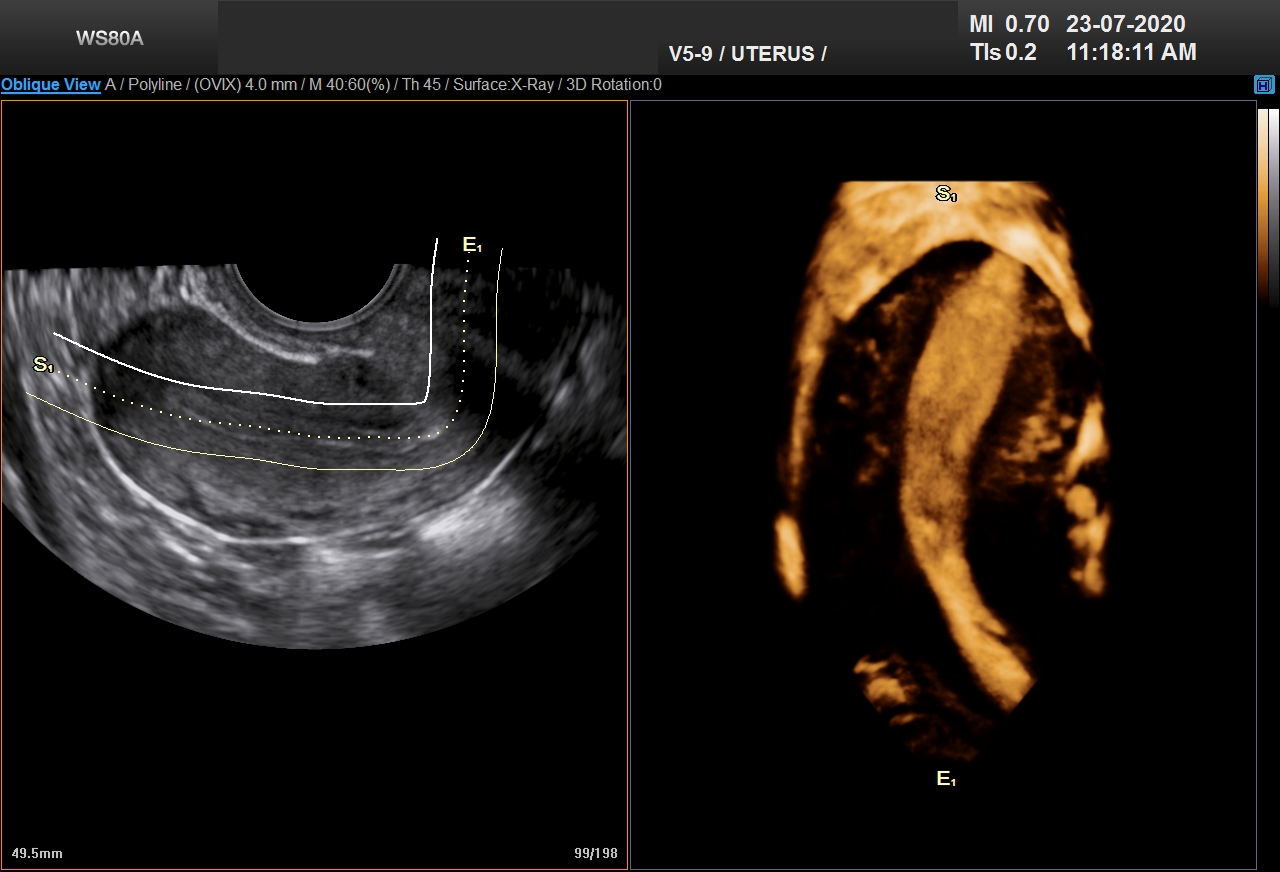
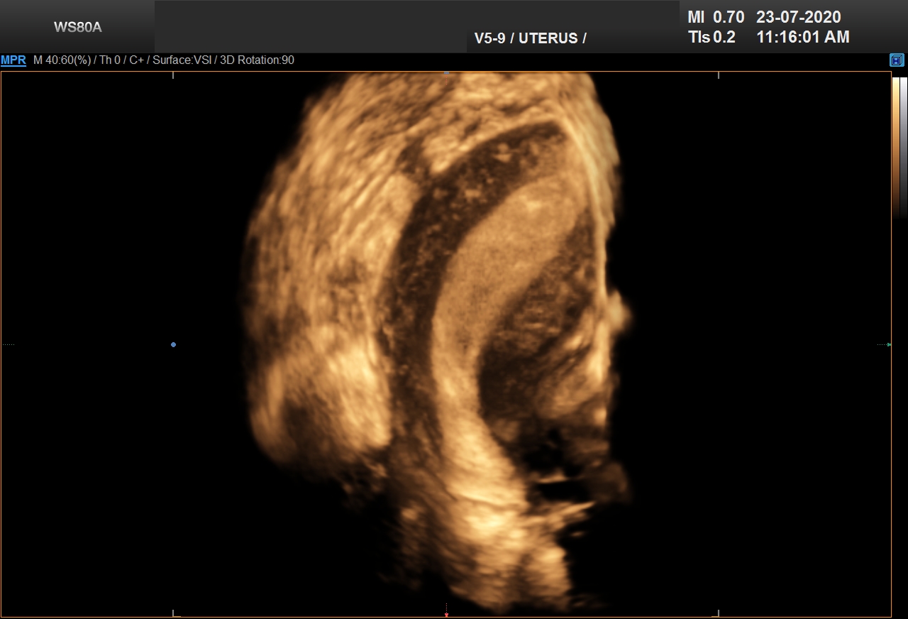
- SAMSUNG WS 80A
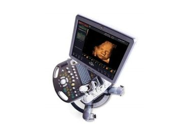
- Voluson S6
- Voluson E8
- Voluson E -10
- Logic 3 PRO Color Doppler (3 units)
Good sonography techniques, updated knowledge, state of the art machines and years of experience are the perfect guide to precise and correct diagnosis. At SFWH, the entire gynaec team and sonology team are adept at diagnosing all sorts of obstetric and gynaec diseases.
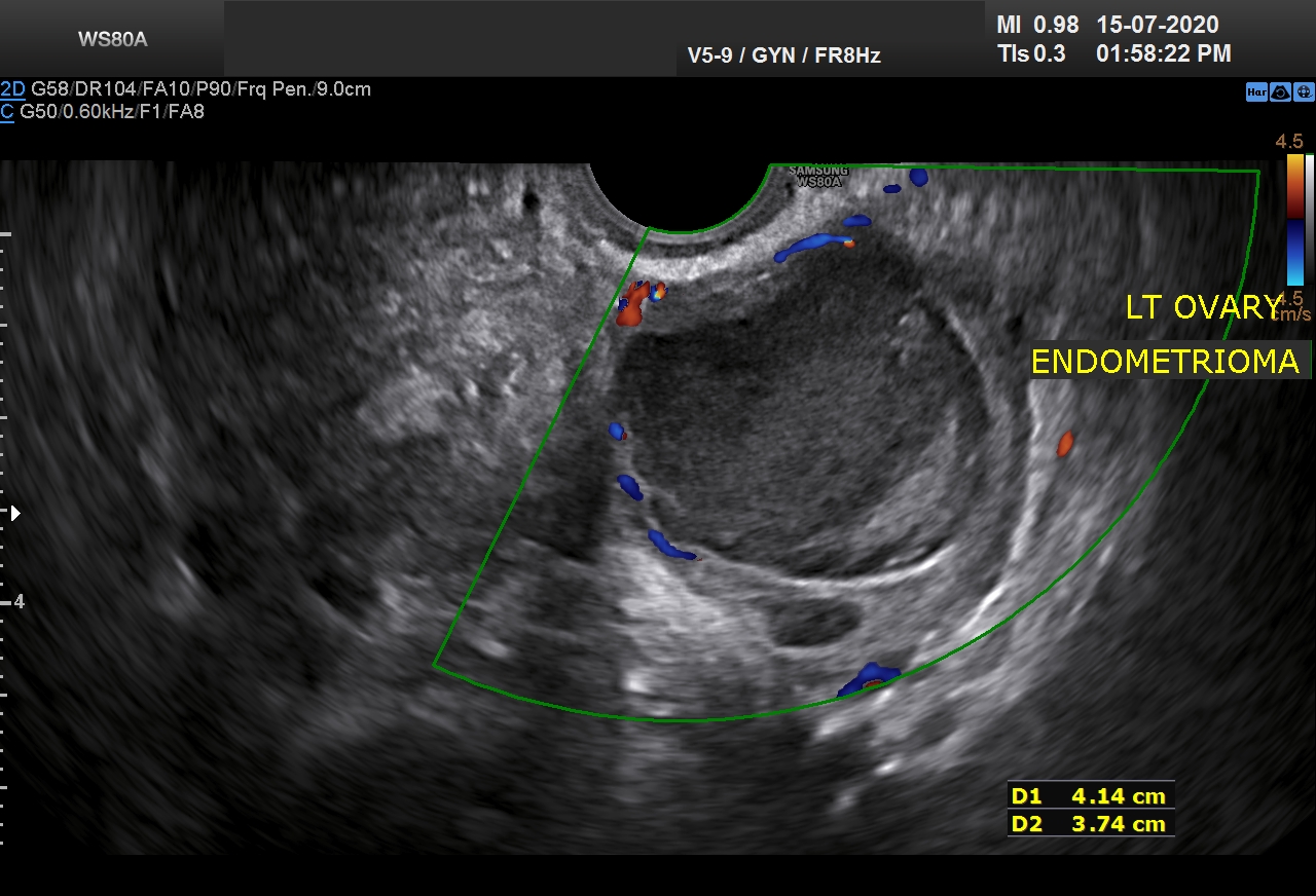
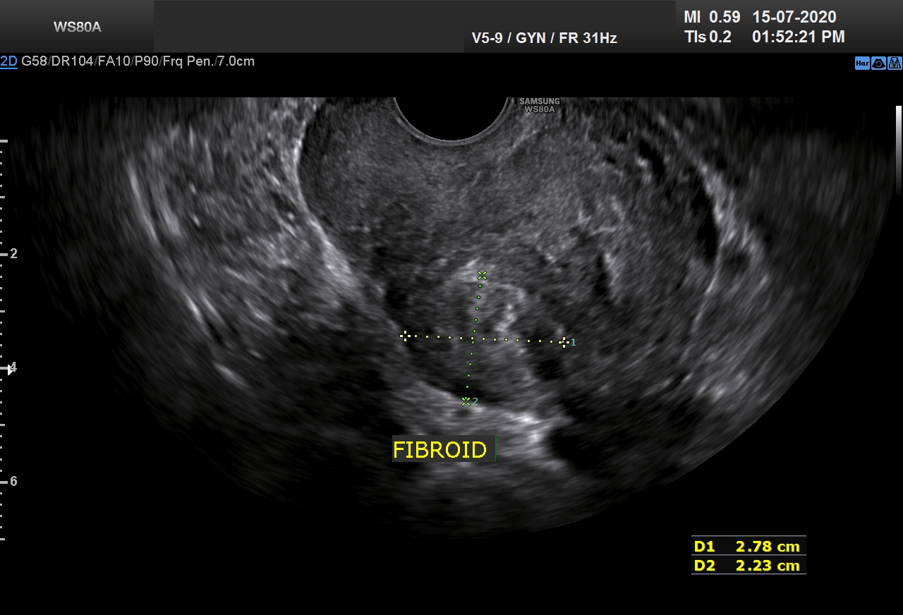
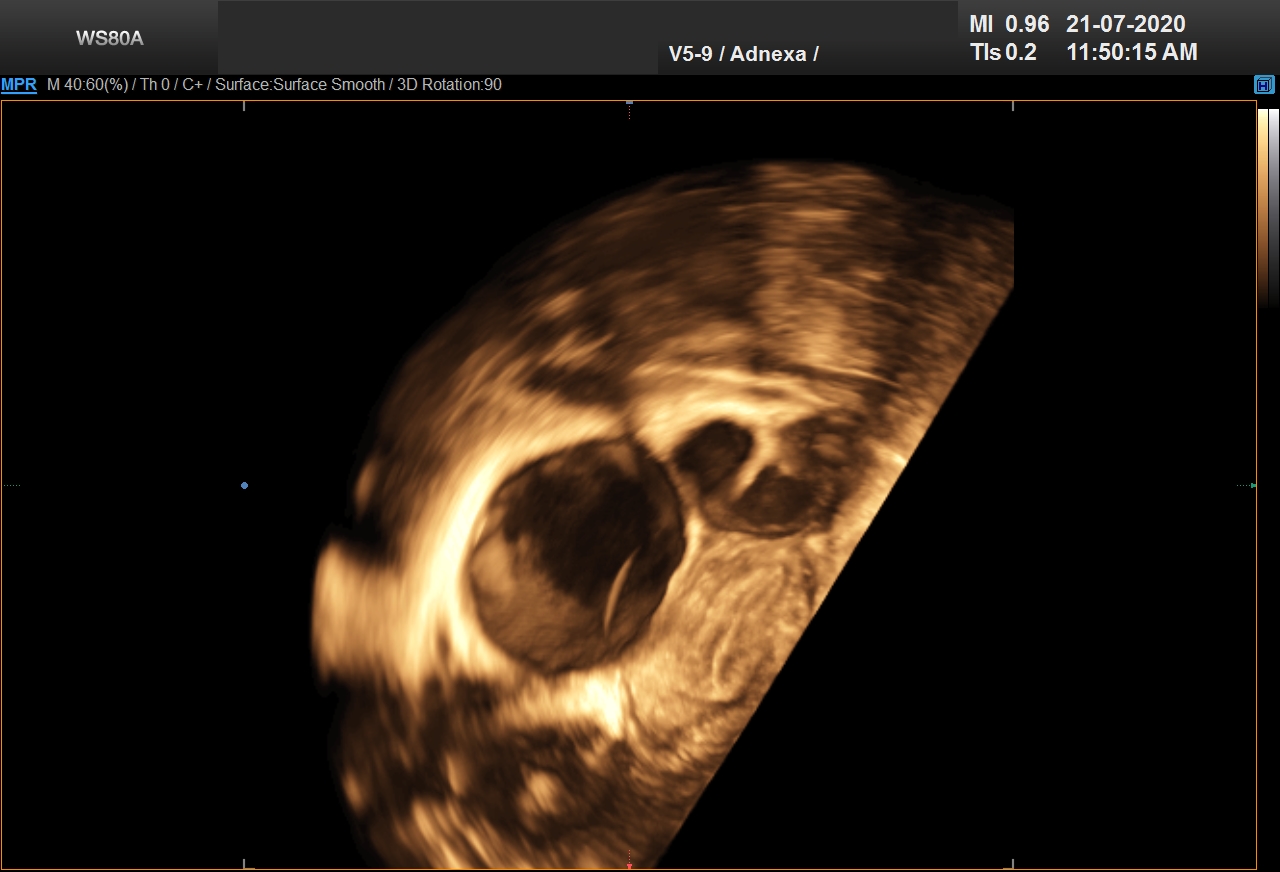
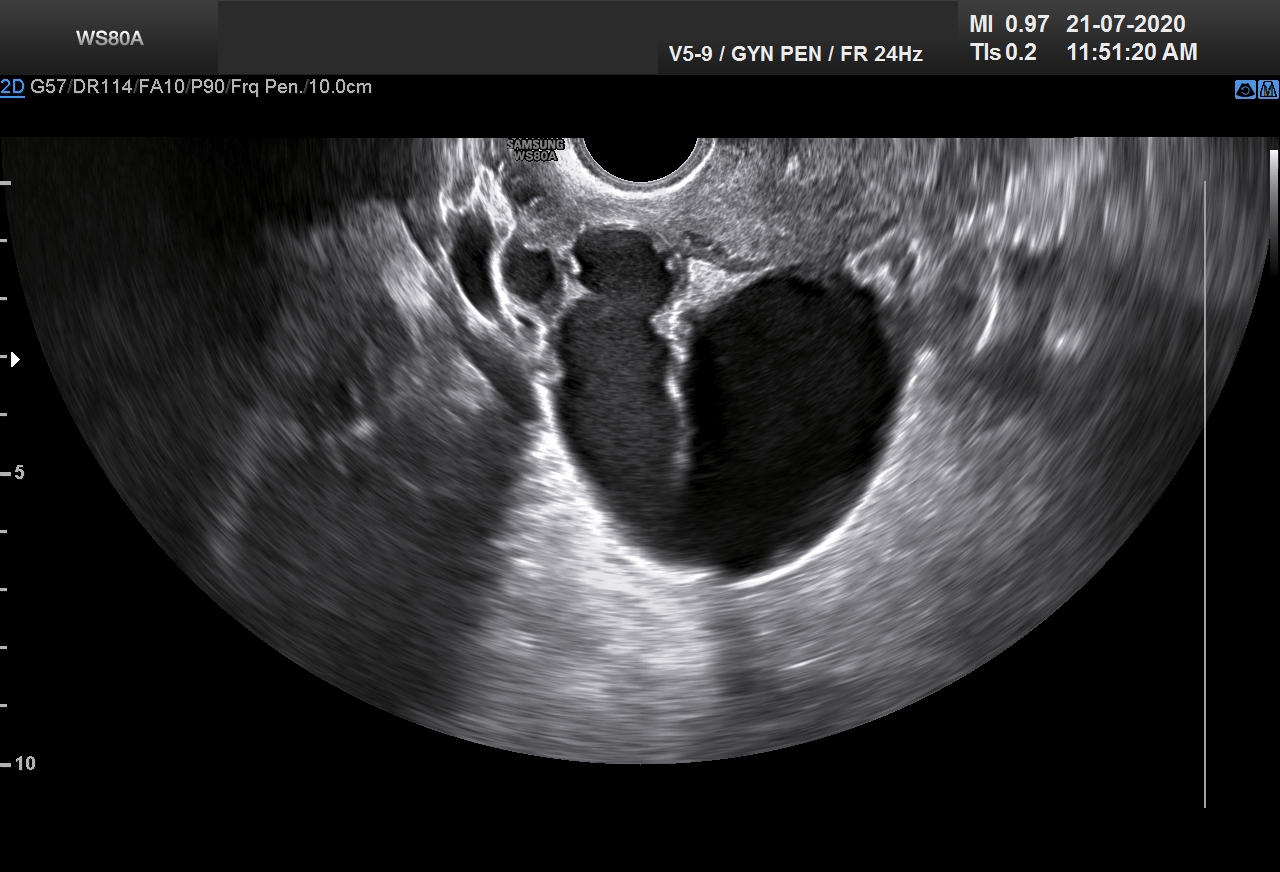
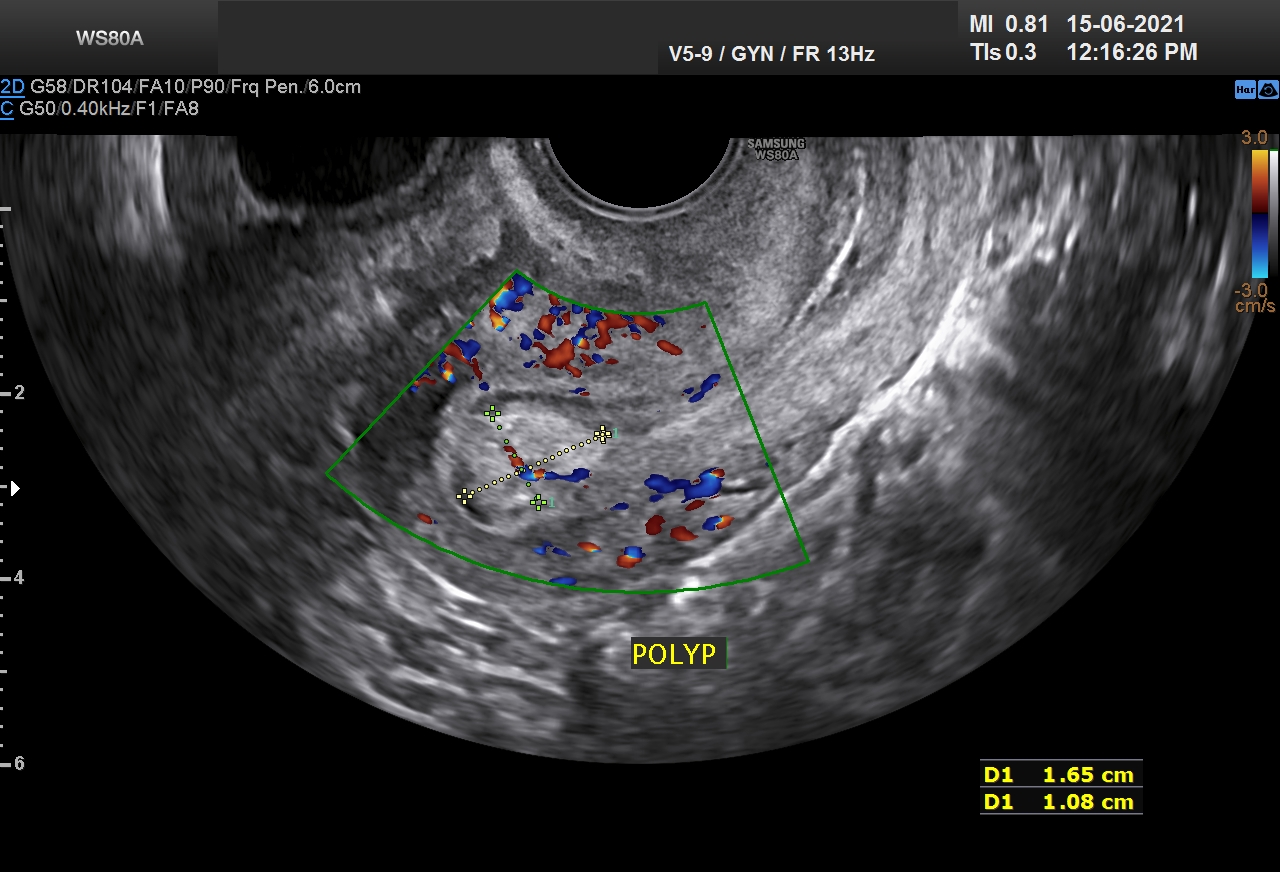
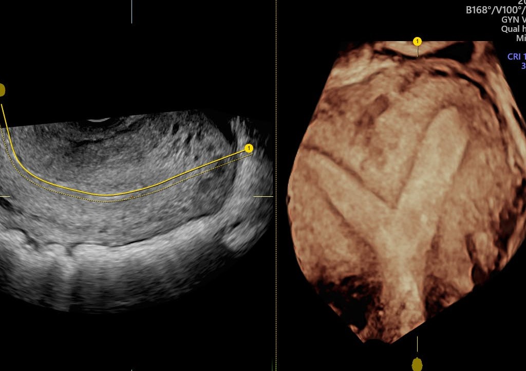
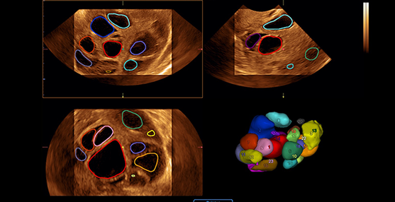
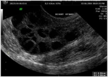
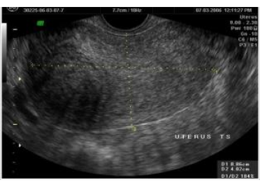
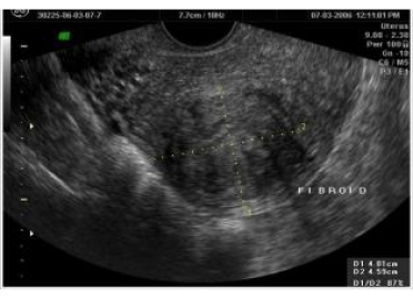
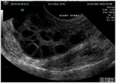
We have full time radiologist Dr Manisha Mandot, Dr.Falguni Parikh visiting sonologist and Dr.Jigish Trivedi and Dr Rakesh Chauhan. They are doing all sorts of obstetric sonography (first trimester, anomaly scan, growth scan and color doppler), and gynaec sonography (follicular study, ovarian volume and mid cycle uterine cavity volume), assesment of uterus, ovary, antral follicle count, endometriotic cysts, dermoid cysts, hydrosalpinx, parovarian cysts, uterine artery doppler, fibroids, adenoma, uterine anomalies like septum, bicornuate, unicornuate uterus.
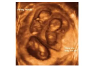
We are doing ultra sound guided procedures like chorion villus sampling (CVS), amniocentesis, selective multifetal reduction (in cases of triplet, quadruplet pregnancies), ovarian cyst aspiration and ovum pick-up.
3D 4D sonography price in Ahmedabad
can be quite overwhelming but not with SFWH. Even our
4D ultrasound price in Ahmedabad
is value for money.
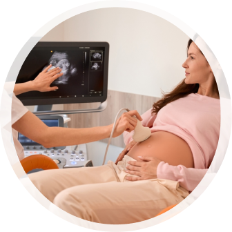
OPD Sonography for Different Diagnosis
Be it Infertility or obstetrics, USG in the basic tool for every Gynaecologist and obstetrician in the outdoor department. It is a noninvasive test used in practically all visits of the patient and gives important clues to the diagnosis of various problems. Early pregnancy diagnosis, number of fetuses, growth and fetal well being, fibroids, ovarian masses, pelvic inflammation, adenomyosis and endometriosis, all sort of basic evaluation can be derived from the first OPD Sonography.
Sonography Advantages
- Because the sonogram uses sound waves and not radiation, it’s completely safe.
- In addition, a sonogram can offer details X-rays can’t.
- It’s painless and in just about every case, the person receiving the sonogram will not be inconvenienced or made to feel uncomfortable in any way.
- Even more important, the sonogram is safe for the unborn child.
Sonography Preparation
There is not too much preparation involved for a sonogram. It is dependent on the area to be examined. For instance, those who are having an abdominal sonogram may be asked not to eat or drink anything for 12 Perior so their doctor can better examine the abdomen. A pregnant woman is usually asked to drink lots of water before her sonogram, as it helps the doctor to see the fetus a little better. Loose, comfortable clothing should be worn in order to make the procedure run a little smoother. Other than that, read all the instructions provided by your doctor.
sunflower success numbers

1500+
IVF and ICSI procedures a year.

21000+
Live births by IVF and ICSI technology.

Life member
AOGS, AICOG, FOGSI, ISAR, ESHRE, ASRM, IFFS, IMA.

Success rate
70% to 76% in IVF ART.
Sonography Instruments
How do 2D Sonography, 3D Sonography, and 4D Sonography ultrasound (USG) examinations differ in pregnant women?
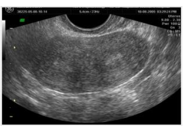
- 2D sonography ultrasound standard examination during pregnancy to determine pregnancy status, the timing of delivery, fetal development, the position of the placenta, and umbilical cord.
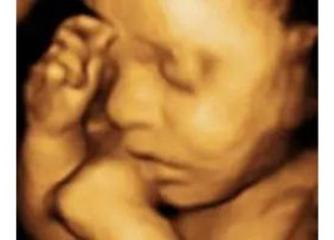
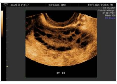
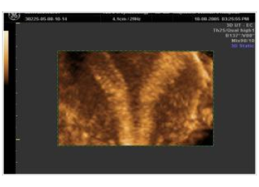
- 3D Sonography Ultrasound Static 3D images that look like pictures of a baby.
- 4D Sonography Ultrasound Animated 3D images that you can view in real-time. Recordings can be saved on different media if you want to have a 4D USG test.
Sonography Segment